

What Is Scintigraphy and How Does It Work?
Scintigraphy is a nuclear medicine examination that images organ function and radiotracer distribution. Small diagnostic activities of tracers, often technetium-99m labelled pharmaceuticals, are used; a gamma camera detects emitted photons.
Planar scintigraphy offers 2D information, whereas SPECT may reconstruct 3D data and SPECT/CT may add anatomical CT. Each organ requires its own tracer and protocol.
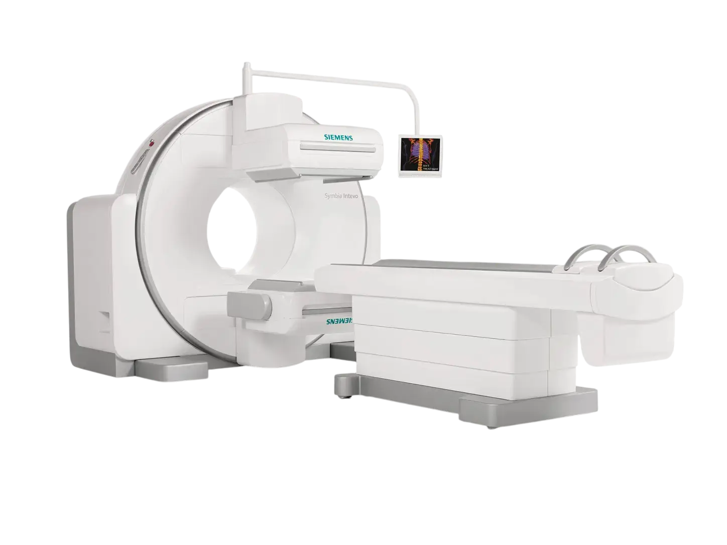
Scintigraphy is primarily diagnostic. Radioiodine and Lu-177 therapies use different indications, doses and precautions.
Why Is Scintigraphy Ordered?
While CT or MRI commonly provides detailed anatomy, scintigraphy may assess perfusion, tracer uptake, drainage or selected metabolic activity; the modalities often complement one another.
Applications include selected assessments of metastatic bone disease, bone infection, kidney function, thyroid nodules, cardiac ischemia, bile flow and lung V/Q mismatch. Imaging findings alone rarely settle every diagnosis.
Types of Scintigraphy: Bone, Thyroid, Renal and Cardiac Scans
Bone, thyroid, MAG3/DTPA/DMSA renal, myocardial perfusion, parathyroid, lung V/Q, HIDA, gastric emptying, lymphoscintigraphy and brain SPECT are distinct investigations.
The table below gives general timing and preparation comparisons. The actual protocol and instructions depend on the selected radiotracer and facility.
| Type | Use | Preparation / timing |
|---|---|---|
| Bone | Metastasis, fracture, infection | Typically hours after injection |
| Thyroid | Nodule function, hyperthyroidism | Iodine/medicine history matters |
| Renal MAG3/DTPA | Function and drainage | Fluids and medicines vary |
| Renal DMSA | Cortex and scarring | Delayed imaging |
| Cardiac perfusion | Ischemia/perfusion | Caffeine/fasting protocol |
| HIDA / gastric | Biliary and gastric emptying | Specific fasting/medicine rules |
Bone Scans for Metastases, Infection and Fractures
A bone scan usually involves IV tracer administration and a wait for uptake in bone. Delayed images often start around two to four hours later; some protocols also obtain early phases.
Increased uptake may reflect metastasis, injury, fracture, arthritis or infection. Correlation with SPECT/CT, CT, MRI or other tests may be required.
Thyroid Scans: Hot and Cold Nodules
Thyroid scanning can characterize functional behavior of nodules and causes of hyperthyroidism. Hot nodules show greater, cold nodules less uptake; neither is by itself a definitive cancer diagnosis.
Recent iodine-based CT contrast, some thyroid medicines and supplements can influence results; decisions about medication changes or delay must be individualized.
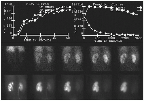
Renal Scintigraphy: MAG3, DTPA, DMSA and Diuretic Renography
MAG3 and DTPA can provide dynamic function or drainage assessment, while DMSA evaluates renal cortical tissue. Diuretic renography assesses suspected obstruction in selected patients.
Renal scintigraphy often answers a different question than stone imaging: how much does each kidney function and how does urine drain? It may complement ultrasound or CT.
Myocardial Perfusion Scintigraphy and Stress Tests
Myocardial perfusion imaging compares perfusion under rest and exercise or pharmacological stress; findings are interpreted with symptoms, ECG and clinical history.
Certain stress agents interact with caffeine. Fasting and medication restrictions vary by protocol. Never stop heart or diabetes medicines without instructions.
Parathyroid, Lung V/Q, HIDA, Gastric Emptying and Lymphoscintigraphy
Parathyroid imaging may localize an overactive gland; V/Q scans assess lung ventilation and perfusion; HIDA evaluates bile passage; gastric emptying studies measure how rapidly a labelled meal leaves the stomach.
Lymphoscintigraphy may guide sentinel node mapping or evaluate lymphatic drainage. The clinical question determines test selection.
How Is Scintigraphy Performed? Step by Step
Before the scan, the team checks the order, pregnancy or breastfeeding, medications and past imaging. The chosen tracer is administered intravenously, orally or by inhalation; uptake and imaging schedules vary.
Patients normally remain still during gamma camera imaging. SPECT/CT adds a short CT scan if indicated. Extra images alone do not mean an adverse finding.
- 1Referral
Confirm clinical indication, medicines and pregnancy/breastfeeding status.
- 2Tracer administration
The tracer is administered IV, orally or by inhalation as indicated.
- 3Uptake interval
Wait the interval required by the organ and tracer.
- 4Imaging
Acquire gamma camera images, sometimes SPECT/CT.
- 5Quality check
Extra images may be needed.
- 6Report
A specialist sends a report to the referring doctor.
How Long Does Scintigraphy Take?
There is no single duration: dynamic renal scans may take tens of minutes, bone scans often mean a visit spanning hours, while some thyroid and specialty scans require more than one imaging time.
Ask about injection time, imaging time, additional imaging and report timing before travelling, especially from outside Ankara.
Preparation: Fasting, Medication, Fluids and Caffeine
Bone and many renal scans often do not require fasting, whereas cardiac stress, HIDA, gastric emptying and certain thyroid protocols require tailored fasting or medicine instructions. Universal rules do not apply.
Provide a list of medicines and supplements, recent iodinated contrast exposure, pregnancy possibility and breastfeeding status. Do not stop prescribed medication without advice.
Risks and Side Effects of Scintigraphy
Scintigraphy uses ionizing radiotracers with a dose depending on the procedure. Serious allergic reactions are rare, with possible injection-site discomfort; stress medicines carry their own risks.
Pregnancy and breastfeeding require tracer-specific advice. Some patients need temporary restrictions on contact or feeding; one universal duration is inappropriate.
Disclose this before scheduling. Only the nuclear medicine team should determine tracer-specific feeding or close-contact restrictions.
What Should You Do After Scintigraphy?
Most people can return to routine after the scan unless advised otherwise. When appropriate, hydration, urination and personal hygiene support tracer clearance; people on fluid restriction should ask for individualized advice.
Follow your written instructions about close contact, breastfeeding, travel and work. Driving restrictions may apply after sedation or selected stress procedures.
When Are Scintigraphy Results Ready?
The specialist reports scan technique, tracer distribution, focal abnormalities and, where appropriate, quantitative measurements. Increased or reduced uptake must be interpreted clinically.
Reporting time depends on workload, extra imaging and scan complexity. Discuss your results with the referring physician.
Scintigraphy vs SPECT/CT, PET/CT, MRI, CT and Ultrasound
Planar scintigraphy yields 2D gamma camera views, SPECT reconstructs 3D function, and SPECT/CT adds localization. PET/CT uses different tracers and detector physics; MRI, CT and ultrasound have distinct anatomical strengths.
The best test depends on disease, clinical question and patient context. Justification and dose optimization matter for radiation-based exams.
| Modality | Main role |
|---|---|
| Planar scan | Functional 2D gamma images |
| SPECT / SPECT-CT | 3D tracer map, optionally localized with CT |
| PET/CT | Positron-emitter molecular data plus CT |
| MRI | Soft tissue detail, no ionizing radiation |
| CT | Fast anatomy using X-rays |
| Ultrasound | Structure/flow using sound, no ionizing radiation |
What Are the Alternatives to Scintigraphy?
Ultrasound/Doppler, MRI, CT, PET/CT, echocardiography, angiography or lab tests may substitute or complement scintigraphy in selected situations, but do not answer all the same functional questions.
Choosing a scan simply because it is newer is not sound; clinical accuracy, the question, radiation exposure and availability matter.
Where Can You Get Scintigraphy in Ankara?
Scintigraphy is carried out by authorized nuclear medicine units. Anka Sağlık Hizmetleri provides a contact channel for enquiries; this page does not claim continuous availability of a specific scan at a particular address.
Patients travelling from neighboring provinces should confirm tracer availability, preparation, delayed imaging schedules, reporting and payment details.
Scintigraphy Prices, Public Coverage and Insurance in Ankara
Fees vary with scan type, radiotracer, protocol, provider and insurance arrangements. Publishing unverified prices would be misleading.
Public and private coverage depends on current rules, clinical indication and contracts. Ask for a written quote with procedure code, potential extras and report delivery details.
Enquire about scintigraphy
State the requested test and referral to ask about preparation, availability and costs. Confirm the site and tracer stock separately.
Contact us →Frequently Asked Questions About Scintigraphy
What is scintigraphy?
Scintigraphy is a nuclear medicine imaging test using a radiotracer and gamma camera to assess organ function and certain tissue changes.
Where is scintigraphy performed in Ankara?
Scintigraphy is performed by authorized nuclear medicine facilities. You may enquire via Anka Sağlık Hizmetleri; verify the actual site, equipment and test availability before attending.
Is scintigraphy a treatment?
Usually no. Scintigraphy is a diagnostic functional imaging test; radioiodine and Lu-177 therapies are separate procedures.
What is a bone scan?
A bone scan evaluates bone metabolic activity; it may help assess selected fractures, infection, metastases or prosthesis concerns.
What does a thyroid scan show?
It shows thyroid tracer distribution and function, particularly in evaluating overactive nodules and causes of hyperthyroidism. It cannot diagnose cancer definitively on its own.
What is renal scintigraphy used for?
It evaluates differential kidney function, perfusion and drainage; MAG3, DTPA and DMSA protocols serve distinct purposes.
What is a MAG3 renal scan?
It is a dynamic renal function and drainage examination; a diuretic may be given when clinically indicated.
What is a DMSA scan?
DMSA evaluates renal cortical tissue and may be used to investigate scarring in selected children.
What is a DTPA scan?
DTPA may be used in selected renal protocols to evaluate perfusion and filtration-related function.
What is myocardial perfusion scintigraphy?
It compares heart-muscle perfusion under rest and stress to assess ischemia and selected coronary artery disease findings.
What is pharmacologic stress testing?
A supervised medicine may simulate stress conditions when exercise testing is unsuitable or not possible.
What is a lung V/Q scan?
It compares lung ventilation and blood flow and may help investigate pulmonary embolism in selected patients.
What is parathyroid scintigraphy?
It may help localize overactive parathyroid tissue in selected patients with hyperparathyroidism; it does not replace biochemical diagnosis.
What is HIDA/hepatobiliary scintigraphy?
It assesses bile movement through the liver, gallbladder and bowel; specific preparation and medicines may be needed.
What is gastric emptying scintigraphy?
It measures emptying of a labelled meal to assess delayed or accelerated gastric emptying.
What is lymphoscintigraphy?
It images lymphatic drainage and may help identify sentinel lymph nodes in selected care pathways.
What is brain SPECT?
It evaluates selected patterns of brain perfusion or function and is not a universal substitute for CT or MRI.
Can scintigraphy diagnose cancer?
Certain scans may reveal metastatic or suspicious areas, but uptake may also reflect infection, injury or healing. Other tests or biopsy may be needed.
How is scintigraphy performed?
The tracer is given by injection, swallowing or inhalation depending on the study; images are acquired with a gamma camera and sometimes SPECT/CT after an appropriate interval.
Is scintigraphy painful?
Imaging is generally painless; IV insertion, lying still or an accompanying stress test may cause discomfort.
How long does scintigraphy take?
Timing varies greatly. Some dynamic scans take tens of minutes; bone scans may require a several-hour uptake period.
When will scintigraphy results be ready?
A nuclear medicine specialist interprets the images; report turnaround depends on facility workflow and complexity.
Do I need to fast before scintigraphy?
Not always. Bone scans typically need no fasting, but cardiac stress, hepatobiliary and gastric-emptying studies may have specific fasting instructions.
Can I drink water before the scan?
Hydration is encouraged for many scans, but some cardiac and gastrointestinal protocols need tailored instructions.
Should I stop my medicines?
Do not stop medication without professional advice. Thyroid medicines, iodine exposures, diuretics and cardiac-stress medicines may require individualized planning.
Does recent iodinated contrast affect a thyroid scan?
Yes. Recent iodinated contrast can affect certain thyroid studies; tell the team when it was given. Any delay depends on the tracer and circumstances.
Should caffeine be avoided before a heart scan?
Caffeine may interfere with certain pharmacologic stress tests. The facility sets a specific avoidance interval; one rule does not fit every protocol.
Can patients with diabetes undergo scintigraphy?
Usually yes, but fasting, medication and insulin timing may need advance coordination, particularly for cardiac and gastric studies.
Can scintigraphy be done during pregnancy?
Pregnancy must be disclosed. The physician evaluates necessity, timing, radiation exposure and alternatives.
Is scintigraphy possible while breastfeeding?
Some examinations are possible, but breastfeeding interruption and safety instructions depend on the radiotracer. Speak with the nuclear medicine team beforehand.
Does scintigraphy involve radiation?
Yes. A radioactive tracer is used. Dose varies by agent, activity, age and any added CT; benefit and risk should be balanced.
Does scintigraphy use less radiation than CT?
There is no universal answer; dose depends on the tracer, activity and CT protocol.
What are scintigraphy side effects?
Serious reactions to radiotracers are uncommon. Injection-site discomfort, rare allergy or stress-drug effects may occur.
What should I do after scintigraphy?
You can often resume normal activities. Follow advice on hydration, urination and any written radiation safety restrictions.
Must I avoid children after a scan?
There is no universal restriction; advice about close contact depends on the radiotracer and dose.
What is SPECT?
Single-photon emission computed tomography collects gamma-camera views around the patient to reconstruct 3D tracer distribution.
What is SPECT/CT?
SPECT/CT combines functional SPECT data with anatomical CT imaging to aid localization when appropriate.
How does scintigraphy differ from PET/CT?
They use different radiotracers and detection principles; PET typically uses positron emitters, while conventional scintigraphy uses gamma cameras.
MRI vs scintigraphy: what is the difference?
MRI uses magnetic fields without ionizing radiation, while scintigraphy uses a radiotracer to evaluate selected organ functions.
Can ultrasound replace scintigraphy?
Sometimes it complements or replaces the scan, but ultrasound and scintigraphy answer different structural and functional questions.
Is a scintigraphy scan the same as radionuclide therapy?
No. A gamma-camera scan is diagnostic; radioiodine or Lu-177 therapy has different purpose, dosing and precautions.
Is scintigraphy covered by Turkish public insurance?
Coverage depends on the examination, indication, facility contracts and current reimbursement rules; confirm before scheduling.
How much does scintigraphy cost in Ankara?
Price varies with scan type, radiotracer and provider. A single unverified quote would be misleading.
Which specialty orders scintigraphy?
A referring specialist orders the scan; the nuclear medicine service performs and interprets it.
Can children undergo scintigraphy?
Yes, when clinically justified; administered activity and preparation are tailored to pediatric patients.
Can scintigraphy be repeated?
It may be repeated when justified; clinicians reassess benefits, prior exposures and alternatives.
What do hot and cold spots mean?
Hot and cold areas indicate greater or reduced tracer uptake; interpretation depends on organ, tracer and context.
Does a positive scintigraphy result mean cancer?
No. Uptake is often nonspecific and may occur with infection, trauma, postoperative change or benign conditions.
How can I arrange a scan from outside Ankara?
If traveling from another province, verify scan type, preparation, waiting time and whether additional-day imaging is required.
Are same-day scintigraphy appointments available?
Not always; radiotracer availability and protocol requirements may require advance scheduling.
References and International Guidance
- Universitat De Barcelona
- Medical University Vienna
- Hôpitaux Universitaires Genève
- LMU University Hospital Munich
- Bukovinian State Medical University
- ACR/RSNA — Nuclear Medicine, General
- ACR/RSNA — Nuclear Medicine Preparation
- ACR/RSNA — Bone Scan
- ACR/RSNA — Renal Scintigraphy
- ACR/RSNA — Thyroid Scan and Uptake
- ACR/RSNA — Safety in Nuclear Medicine
This is general education. A physician must determine indication, preparation and interpretation.