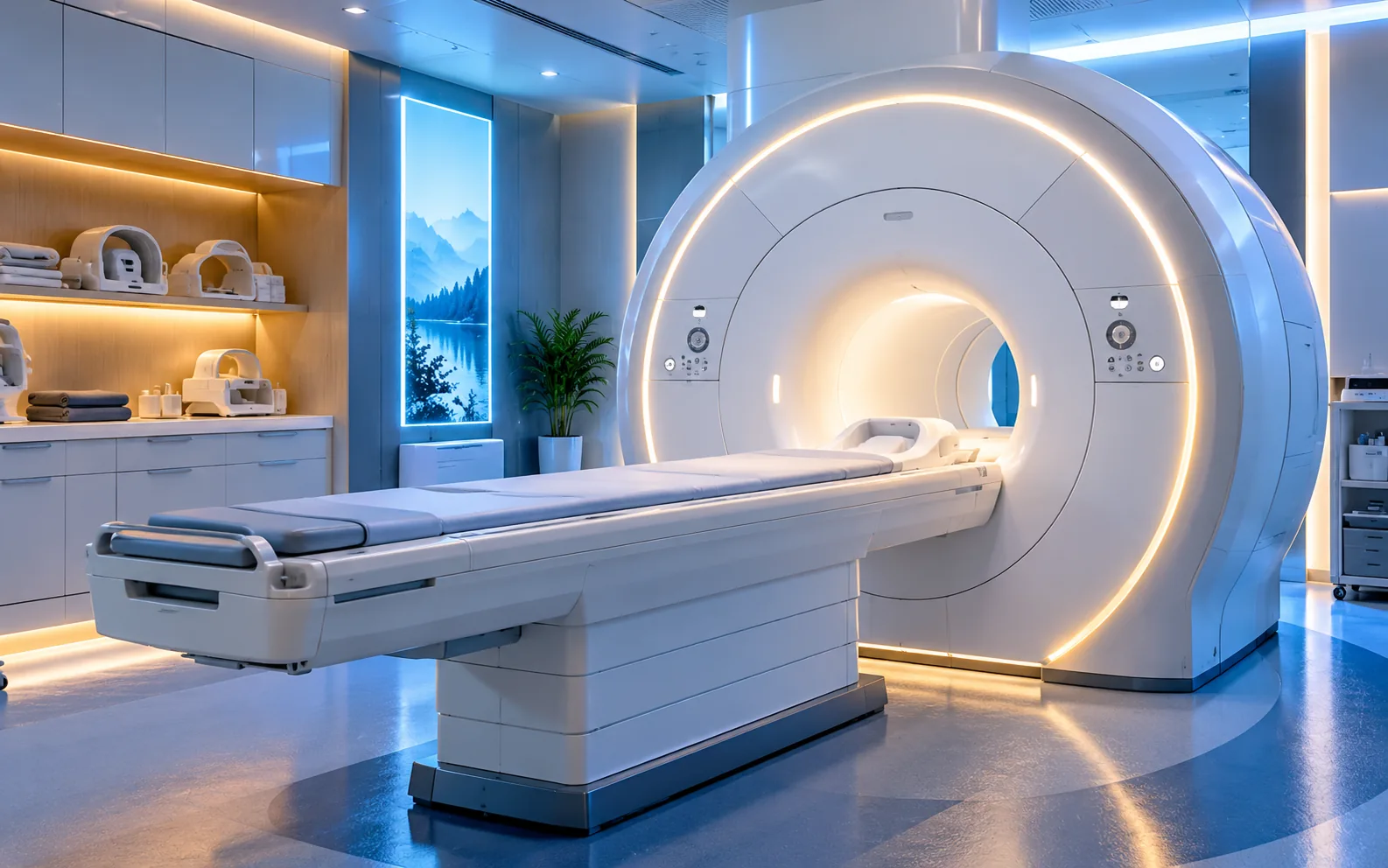
What Is 3 Tesla MRI?
Magnetic resonance imaging (MRI) uses a strong magnet, radiofrequency pulses and a computer to generate detailed images of tissues and organs. 3 Tesla MRI describes a system with a 3-tesla main magnetic field; “3T MRI scan” refers to the examination performed on such a system.
With appropriate protocols, 3T scanners can use a higher signal-to-noise ratio to depict fine anatomical detail. Diagnostic value also depends on the anatomy, clinical question, hardware, coils, patient motion and radiology protocol. Higher field strength is not automatically best for every patient.
3 Tesla MRI is an imaging test, not a treatment. “3T” is a technical capability, not a guarantee of superior results; indication and protocol matter.
3 Tesla vs 1.5 Tesla MRI
The main difference is magnetic field strength. Both systems are used clinically, and choosing between them depends on the examination and the patient’s safety profile.
| Feature | 1.5 Tesla MR | 3 Tesla MR |
|---|---|---|
| Magnetic field | 1.5 T | 3 T |
| Signal potential | Sufficient for many routine studies | Higher signal in suitable protocols |
| Image detail | High clinical image quality | Potentially finer detail for some exams |
| Scan duration | Protocol-dependent | May be shorter for some sequences; not guaranteed |
| Metal and artifacts | May be preferred with certain implants/metal | Some artifacts and restrictions can increase |
| Who is it for? | Clinical need, scanner capability and implant conditions determine selection. | |
Evidence: Investigative Radiology review (2021) and FDA MRI safety guidance.
Benefits and Limitations of 3T MRI
Potential benefits in selected patients
- Higher signal may support thinner slices or higher spatial resolution.
- Can support specialized imaging of the brain, selected joints and prostate.
- May offer flexibility in the balance of scan speed and detail.
- MRI does not use ionizing radiation.
Important limitations
- Metal-related image distortion and some artifacts can become more prominent at higher field strengths.
- Radiofrequency heating and implant restrictions require careful safety checks.
- Implanted and external devices must be evaluated against their specific MRI conditions.
- A 3T scan is not invariably faster, more accurate or contrast-free.
Which Body Areas Can Be Examined With 3T MRI?
3T MRI can be applied to many regions, but specialized studies need appropriate coils, software, protocols and trained teams. These are general medical applications and do not imply that every protocol is necessarily offered by Anka.
Whole-body MRI is not a routine cancer screening test for everyone. Broad scans in asymptomatic people may lead to incidental findings and unnecessary follow-up. The scope should be clinically guided.
How Is a 3T MRI Scan Performed?
- 1Referral and protocolThe clinical question is reviewed to decide the examination, field strength and contrast need.
- 2MRI safety screeningImplants, surgery, pregnancy possibility, kidney issues and metallic foreign bodies are reviewed.
- 3PreparationMetal items are removed; an appropriate gown and hearing protection are provided as needed.
- 4ScanningA coil is positioned and the table moves into the scanner. Remaining still is important; contrast is administered if indicated.
- 5Review and reportImages are checked, interpreted by a radiologist and reported through the provider’s process.
Rhythmic knocking and tapping noises are expected. The staff can communicate with the patient. Remaining still matters, and some abdominal or cardiac scans require brief breath holds.
Preparing for a 3T MRI
- Bring your referral and prior relevant imaging or reports.
- Disclose pacemakers, neurostimulators, cochlear implants, clips, stents, pumps or fragments; bring implant documentation.
- Tell the team about possible pregnancy, significant kidney disease, prior contrast reaction or claustrophobia.
- Remove jewelry, watches, belts, clips, piercings and electronics. Follow clothing instructions for possible metal fibers.
- Fasting depends on the examination; follow site-specific instructions for abdominal exams, sedation and special contrast protocols.
- Do not stop routine medications on your own; discuss questions with your clinicians.
Contrast-Enhanced vs Non-Contrast 3T MRI
Contrast-enhanced MRI commonly uses an intravenous gadolinium-based agent, which differs from the iodinated agents used in many CT exams. Contrast may be indicated for selected tumor, inflammatory or vascular questions, but is not necessary for all MRI studies.
Previous reactions, renal function, pregnancy and repeated exposure affect contrast decisions. Rare reactions and special risks in severe renal disease require clinical evaluation.
Is 3T MRI Safe? Who Needs Special Screening?
MRI does not use ionizing radiation but involves a powerful static magnetic field, changing gradients and radiofrequency energy. Safety screening addresses metal objects, implanted devices, possible heating and hearing protection.
Do not bring phones, oxygen tanks or unapproved metal into the scanner room—even when imaging is not underway. Follow the MRI team’s screening instructions.
Situations requiring special assessment
- Pacemakers, ICDs, cochlear implants, neurostimulators, pumps and some vascular clips
- Possible metallic foreign bodies, especially near the eyes, or unknown implants
- Pregnancy, pediatric exams, sedation needs or substantial claustrophobia
- Significant kidney disease or prior contrast reactions if contrast is planned
A device being eligible at 1.5T does not automatically mean it is eligible at 3T. Manufacturer-specified MR Conditional requirements—including field strength—must be verified.
What Happens After the Scan?
Images are checked for technical quality and interpreted by a radiologist. Ask how to receive images, reports and comparisons with earlier scans. Most patients can resume normal activities without sedation; follow individual instructions after contrast or sedation.
3T MRI in Ankara: Appointments, Costs and Nearby Provinces
For people searching for 3T MRI services in Ankara, the key is to match the examination, scanner and safety protocol to the medical indication. When contacting Anka Sağlık Hizmetleri, share the body area, referral, contrast needs and relevant implant information.
If traveling from Kırıkkale, Çankırı, Eskişehir, Bolu, Konya, Kırşehir or nearby provinces, confirm the exact location, suitability of the 3T protocol, appointment, reporting and current fee before traveling. Insurance conditions and prices vary; no unverified pricing or guarantees are stated here.
Ask About 3 Tesla MRI
Contact the team to clarify the requested exam, clinical referral, safety conditions and appointment options. This page does not itself guarantee a booking or a specific 3T scanner.
Appointment / Information Form☎ 0538 350 74 45Frequently Asked Questions About 3T MRI
Answers to common questions before and after 3T MRI. Individual medical decisions require a clinician’s assessment.
What is a 3 Tesla MRI?
3 Tesla MRI is imaging performed using an MRI scanner with a 3-tesla magnetic field. Unlike CT, MRI does not use ionizing X-rays.
What is the difference between 3T and 1.5T MRI?
A 3T system has twice the static magnetic field of a 1.5T system. Suitable protocols may deliver more signal, finer detail or faster acquisition, but 3T is not always better; implants and artifacts sometimes favor 1.5T.
How long does a 3T MRI scan take?
Many MRI exams take around 15–45 minutes of scanning, but some require 60 minutes or more depending on the anatomy, sequences, contrast and motion. Preparation time can be additional.
Does 3T MRI provide clearer images?
In some brain and musculoskeletal applications, increased signal can support greater detail. Image quality still depends on coils, protocols, patient motion and interpretation, not field strength alone.
Does 3 Tesla MRI use radiation?
No. MRI uses magnetic fields and radiofrequency waves, not the ionizing X-rays used in CT or radiography. MR-specific safety screening is still essential.
Is a 3T MRI painful?
It is usually painless. Staying still can be uncomfortable; contrast-enhanced scans may involve an IV needle, and the scanner makes loud noises.
Is a 3T MRI always performed with contrast?
No. Many 3T studies are non-contrast. The need for intravenous gadolinium contrast depends on the clinical indication and radiology protocol, not field strength alone.
Can patients with kidney disease undergo contrast-enhanced 3T MRI?
Tell your care team about kidney disease. A clinician considers the gadolinium agent, renal function and alternatives, and may request recent kidney-function results.
Can someone with a pacemaker or metal implant have a 3T MRI?
Eligibility depends on the exact device and its MR Safe, MR Conditional or MR Unsafe labeling. Some devices are limited to specified field strengths or operating conditions. Bring device information for screening.
What if I have orthopedic or dental implants?
Many modern implants can be evaluated for MRI, but no implant should be assumed safe without appropriate checks. Device type, location, labeling and potential image artifacts matter.
Can a 3T MRI be performed during pregnancy?
MRI does not use ionizing radiation. During pregnancy, clinical need and field strength require professional assessment, while gadolinium is generally avoided unless essential. Always disclose pregnancy.
Can breastfeeding patients have contrast-enhanced 3T MRI?
Inform the radiology team if you are breastfeeding. Discuss the particular contrast agent and current guidance; a routine interruption is not automatically necessary for everyone.
Is 3T MRI an open MRI?
3T describes magnetic field strength, whereas open/closed describes the scanner design. Many clinical 3T units are tunnel-style systems; ask about bore size and options if you have claustrophobia.
What if I am claustrophobic?
Tell the team in advance. Communication, hearing protection and positioning may help. A clinician can assess sedation or an alternative scanner; transportation arrangements may be needed after sedation.
Why might a 3T brain MRI be requested?
Appropriate MRI protocols can help evaluate brain, pituitary, seizure-related, tumor, vascular and demyelinating conditions as clinically indicated. The referring clinician selects the examination.
Can 3T prostate MRI diagnose cancer with certainty?
Multiparametric prostate MRI may support lesion assessment and biopsy planning but does not establish cancer definitively by itself. Results are integrated with clinical findings and, when indicated, pathology.
Do I need to fast before a 3T MRI?
Fasting is not routinely required for many brain and joint exams. Abdominal, pelvic, sedation or some contrast protocols may require preparation. Follow your facility’s specific instructions.
Must I remove jewelry and clothing?
Jewelry, metal accessories and removable electronic items are not taken into the MRI area. Some fabrics contain metallic fibers, so a facility may provide MRI-safe clothing.
How much does a 3T MRI cost in Ankara?
Prices vary by body region, contrast, protocols, reporting and coverage. No unverified price is published here; contact Anka Sağlık Hizmetleri for current details.
Does public or private insurance cover a 3T MRI?
Coverage depends on authorization, medical indication, provider arrangements and policy terms. Confirm directly with your insurer and the facility.
When will I receive my 3T MRI report?
Reporting times depend on examination complexity and the provider’s workflow. Ask for the expected report and image delivery process when booking.
Can children undergo 3T MRI?
Pediatric MRI depends on clinical need, age, ability to remain still and possible sedation. Appropriate protocols and screening are essential.
Can I resume normal activities after 3T MRI?
Most people can resume normal activities after an MRI without sedation unless otherwise instructed. Follow individual instructions after sedation, contrast reactions or added procedures.
Can patients outside Ankara request 3T MRI information?
People from nearby provinces can request information. Confirm the referral, 3T availability, imaging protocol, travel, pricing and appointment terms before traveling.
International Medical References
- U.S. Food and Drug Administration (FDA) — MRI: Technology and General Overview.
- U.S. Food and Drug Administration (FDA) — Benefits and Risks of MRI.
- American College of Radiology / RSNA — MRI Safety.
- American College of Radiology / RSNA — Body MRI (2026).
- American College of Radiology / RSNA — MRI During Pregnancy.
- American College of Radiology / RSNA — Contrast Material.
- Investigative Radiology — 1.5 vs 3 Tesla MRI: A Review of Favorite Clinical Applications (2021).
- American College of Radiology — MR Safety Manual (2026).