
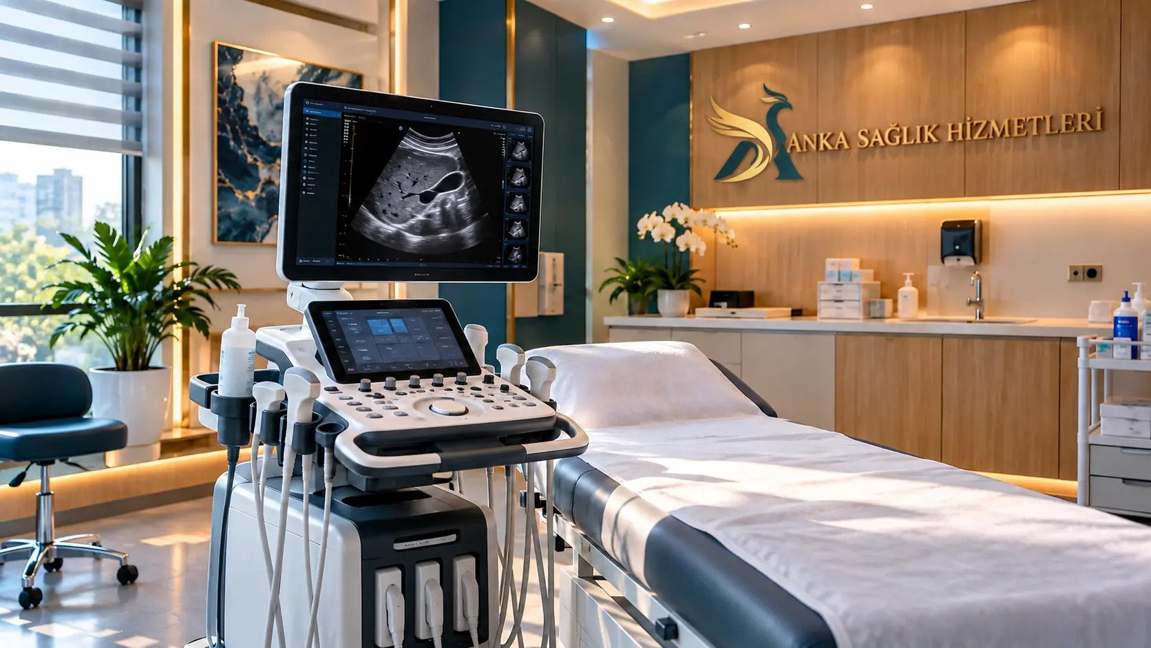
What Is Ultrasound (Sonography / USG)?
Sonography creates real-time images using high-frequency sound waves reflected from tissues. Ultrasound, sonography and USG describe the same underlying technology. A handheld transducer sends sound waves and receives echoes that are converted into images.
Ultrasound uses no ionizing radiation. It is usually quick and noninvasive, but it cannot definitively characterize every organ or finding.
How Does an Ultrasound Machine Work?
An ultrasound machine includes a transducer, display and processing console. Water-based gel removes air between the probe and skin so sound waves can pass more effectively. Echoes form 2D images; Doppler adds blood-flow assessment and elastography can assess stiffness in selected tissues.
Because images are obtained in real time, movement of tendons, blood flow and fetal movement may be assessed as clinically appropriate. Bowel gas, depth and bone can limit visualization.
Why Might a Clinician Request an Ultrasound?
An ultrasound referral does not itself mean a diagnosis. It can help investigate symptoms, monitor findings or inform treatment decisions. The clinician selects the examination in view of symptoms and other tests.
- Investigation of abdominal pain, gallstones and selected liver findings.
- Assessment of kidney, urinary tract and bladder concerns.
- Assessment of thyroid nodules, neck lumps and superficial swellings.
- Investigation of breast lumps and cystic versus solid features alongside other tests.
- Doppler assessment of suspected blood-flow problems, stenosis or venous thrombosis.
- Monitoring pregnancy and fetal development at appropriate stages.
- Assessment of musculoskeletal and soft-tissue complaints.
- Imaging guidance for selected biopsies and fluid aspirations.
Ultrasound Types and Body Areas
Ultrasound examinations differ by probe, depth, settings and protocol. The applications below describe medical examination types; confirm whether a particular service is available through Anka Sağlık Hizmetleri before booking.
What Is Doppler and Color Doppler Ultrasound?
Doppler analyzes shifts in reflected sound associated with moving blood cells. Color Doppler maps flow information over images, while spectral Doppler displays flow-related measurements in graphs. It is used for venous, carotid, renal and other selected vascular examinations.
How Is an Ultrasound Scan Performed?
- 1Referral and preparationThe requested body region and any fasting or bladder instructions are confirmed.
- 2PositioningYou lie or sit in the position required for the examination.
- 3Gel and transducerGel is placed on the skin and the probe is moved to acquire real-time images.
- 4Image assessmentMeasurements, Doppler views where needed and images are recorded.
- 5ReportingGel is wiped off and an authorized clinician interprets images and reports findings.
External ultrasound often takes about 10–30 minutes, with longer examinations for more detailed studies. Internal transvaginal or transrectal examinations follow additional preparation and consent steps.
Preparing for Ultrasound: Fasting and Bladder Filling
Incorrect preparation can reduce image quality or require a repeat visit. The information below is general; the exact fasting time and amount of water should come from the imaging facility.
Upper abdomen / gallbladder
Some protocols may request about 6–8 hours of fasting. Ask for tailored advice in pregnancy, diabetes, childhood or with medication.
Pelvic / lower abdomen
A full bladder may be requested; the amount and timing of water depend on local instructions.
Transvaginal ultrasound
An empty bladder is commonly requested. Explanation and consent are essential.
Thyroid, breast and soft tissue
Fasting is usually unnecessary. Wear clothing allowing convenient access to the area.
Doppler ultrasound
Preparation varies with the vessels examined; leg and abdominal Doppler instructions may differ.
Pediatric ultrasound
Instructions should be age- and examination-specific rather than copied from adult fasting protocols.
Do not discontinue prescribed medicine without clinical advice. Bring your referral and relevant prior imaging reports when available.
Ultrasound in Pregnancy, Detailed Anatomy and 3D/4D Scans
Obstetric ultrasound evaluates pregnancy location, dating, fetal development and anatomy at appropriate stages. A detailed anatomy scan has different aims from routine follow-up and is organized by the obstetric team.
2D creates planar views, 3D volume views, and 4D moving volume views. A more elaborate image does not automatically mean better diagnosis. FDA and AIUM stress medically justified, appropriately trained and prudent use in pregnancy.
Diagnostic ultrasound does not use X-rays, but prolonged nonmedical keepsake scans and unnecessary Doppler exposure are discouraged.
Does Contrast-Enhanced Ultrasound (CEUS) Exist?
Most routine ultrasound exams do not require an injected contrast agent. Contrast-enhanced ultrasound (CEUS) is a specialized technique that uses IV microbubble agents to assess tissue perfusion in selected cases. These agents differ from iodinated CT or gadolinium MRI contrasts.
CEUS is not offered everywhere. A clinician assesses indications and possible adverse reactions. If a referral says contrast ultrasound, confirm the exact procedure.
Ultrasound Safety, Risks and Side Effects
Standard diagnostic ultrasound carries no ionizing-radiation exposure and has a strong safety record in appropriate medical practice. Acoustic energy may cause thermal or mechanical effects, so AIUM recommends the ALARA principle.
- Probe pressure can cause temporary tenderness, particularly in painful areas.
- Internal examinations may cause transient discomfort and require informed consent.
- Biopsy or other ultrasound-guided procedures carry risks distinct from standard imaging.
- Specialized contrast agents require additional safety assessment.
- Unnecessary prolonged or repeated nonmedical scanning should be avoided.
Benefits and Limitations of Ultrasound
Benefits
No ionizing radiation, real-time imaging, usually efficient examinations, dynamic assessment and guidance for some procedures.
Limitations
Gas, bone, depth and body habitus may restrict imaging. Results are operator-dependent, and some findings require MRI, CT, endoscopy or pathology.
Ultrasound vs MRI, CT and X-ray
Ultrasound, MRI and CT answer different diagnostic questions. Choosing another test solely for convenience or cost may not be appropriate.
| Method | How it works | Typical uses / limits |
|---|---|---|
| Ultrasound | Sound waves; no X-rays | Abdomen, superficial structures, Doppler and pregnancy; gas and bone can limit |
| MRI | Magnetic fields and RF; no X-rays | Brain, spine, joints and selected soft tissue; implant screening and longer exams |
| CT | Ionizing X-rays | Fast assessment in selected emergencies, trauma, lung, bone and deep abdomen |
| X-ray | Ionizing X-rays | Bone and selected chest imaging; less soft-tissue detail |
Color Doppler is not a separate radiation-based modality; it is an ultrasound technique for blood-flow assessment.
After Your Ultrasound Scan
After a standard scan, the gel is wiped away and most patients can resume daily activities. Medical reporting is separate from image acquisition. Results are interpreted alongside symptoms and other tests. Special post-procedure instructions take priority after internal examinations, sedation, biopsy or contrast use.
Understanding Common Ultrasound Report Terms
- Cyst: a fluid-containing structure; significance depends on appearance and organ.
- Solid lesion or nodule: tissue-like finding, not automatically malignant.
- Hypoechoic / hyperechoic: darker or brighter ultrasound appearance relative to adjacent tissue.
- Hydronephrosis: dilation of the kidney collecting system, sometimes requiring investigation.
- Possible steatosis: liver appearance that may be consistent with fat accumulation; assess with clinical/laboratory findings.
- Doppler flow findings: visualization and measurements of flow; isolated measurements do not stand alone as a diagnosis.
Do not infer a definitive diagnosis from a single report term. Discuss the radiologist’s impression and recommendations with your referring clinician.
Where to Get an Ultrasound in Ankara: Appointments and Prices
In Ankara, ultrasound is provided by appropriately authorized hospitals, imaging centers and specialty services. Before seeking an appointment, confirm exactly which exam has been prescribed.
You can contact Anka Sağlık Hizmetleri about ultrasound and other imaging examinations. Confirm the actual location, whether the requested subtype is available, the appointment and any fees. This page does not claim a specific machine, specialist or examination location.
If traveling from Kırıkkale, Çankırı, Kırşehir, Bolu, Eskişehir, Konya or nearby provinces, confirm preparation, report delivery and referral acceptance before travel. Contact the provider to check fees and insurance conditions.
Ultrasound Information and Appointment Request
Share the exact exam request, prior reports and any preparation questions to confirm available scanning, the facility, charges and scheduling.
Contact / Appointment Form☎ 0538 350 74 45Frequently Asked Questions About Ultrasound
Find answers about ultrasound time, fasting, bladder filling, Doppler, pregnancy, reports, fees and booking in Ankara. This is general medical information, not personalized advice.
What is ultrasound and what is it used for?
Ultrasound (sonography) uses high-frequency sound waves to create images of organs and tissues. It is used for abdominal organs, thyroid, breast, musculoskeletal structures, blood vessels and pregnancy when clinically indicated.
Does ultrasound use radiation?
Ultrasound uses sound waves rather than the ionizing X-rays used in radiography or CT. It should still be used appropriately for a medical purpose.
How long does an ultrasound take in Ankara?
Many routine examinations take about 10–30 minutes. Doppler, detailed pregnancy imaging and complex procedures may take 45–60 minutes or longer, not including preparation or reporting.
Does an ultrasound hurt?
External ultrasound is usually painless. Pressure over a tender area, a full bladder or an internal scan can cause discomfort; tell the operator if it hurts.
Do I need to fast before an ultrasound?
Not every scan requires fasting. Upper abdominal and gallbladder ultrasound may require fasting, while thyroid, breast and many soft-tissue scans usually do not. Follow examination-specific instructions.
Why might I need to drink water?
A full bladder can improve the view during selected pelvic and lower-abdominal exams. Your facility may ask you to drink water and avoid urinating; it is not required for every ultrasound.
Can I use the bathroom before my scan?
You may be asked to keep your bladder full for transabdominal pelvic imaging. Transvaginal ultrasound often requires an empty bladder. Follow your individual instructions.
What is a complete abdominal ultrasound?
A complete abdominal ultrasound evaluates abdominal and sometimes pelvic structures according to the referral and protocol. The exact organs included and need for bladder filling vary.
Which organs are examined in an upper abdominal ultrasound?
It commonly examines the liver, gallbladder, biliary system, visible pancreas, spleen, kidneys and sometimes major vessels. Bowel gas may limit some views.
Are lower abdominal and pelvic ultrasound the same?
The terms may overlap in everyday use. Pelvic ultrasound evaluates the bladder, uterus, ovaries and other pelvic structures as clinically indicated; the referral determines the scope.
What is Doppler ultrasound?
Doppler is an ultrasound technique that assesses blood flow direction and velocity. Color, spectral and power Doppler can be used in selected vascular assessments.
How is color Doppler different from standard ultrasound?
B-mode ultrasound shows tissue structure; color Doppler overlays flow information. It is a form of ultrasound rather than a different radiation-based test.
What does thyroid ultrasound show?
It assesses thyroid size, texture, nodules, cysts and sometimes neck lymph nodes. Ultrasound appearance alone does not establish cancer; risk stratification and biopsy decisions may be needed.
Does breast ultrasound replace mammography?
Not routinely. Breast ultrasound can complement other imaging for a lump or cyst; mammography or MRI may still be needed depending on age, risk and clinical findings.
Can ultrasound show kidney stones?
Some kidney stones and urinary obstruction can be detected, but small stones and some ureteric stones may be missed. CT or another test may be needed.
Is ultrasound safe during pregnancy?
Diagnostic obstetric ultrasound is generally considered safe when used for a medical reason by trained personnel with prudent exposure. Prolonged nonmedical keepsake scans are discouraged.
Is a routine pregnancy scan the same as a detailed anatomy scan?
Not exactly. Routine obstetric scans and detailed fetal anatomy examinations have different aims and protocols. Timing and referral depend on obstetric care.
How is transvaginal ultrasound performed?
When appropriate and with consent, a covered, lubricated probe is gently placed in the vagina for closer views of the uterus and ovaries. The patient may request stopping the procedure.
Can ultrasound be performed during menstruation?
Some scans can be performed during menstruation, but optimal timing depends on the clinical question. Discuss comfort and any preparation with the team.
Can ultrasound definitively diagnose cancer?
Ultrasound may reveal or monitor suspicious lesions but generally does not confirm cancer by itself. Further imaging and tissue sampling may be needed.
Can ultrasound examine the stomach and intestines?
Specialized bowel ultrasound is possible, but gas limits imaging of the stomach and bowel. Endoscopy, CT or MRI may be more suitable for some questions.
What is the difference between ultrasound and MRI?
Ultrasound uses real-time sound waves; MRI uses a magnetic field and radiofrequency energy and can offer different soft-tissue detail. Neither uses ionizing X-rays; the choice depends on the question.
How does ultrasound differ from CT?
Ultrasound uses sound waves without ionizing radiation; CT uses X-rays and can be more appropriate for certain emergency, lung, bone, bowel or deep-organ questions.
What do 3D and 4D ultrasound mean?
2D shows planar views, 3D produces volume images, and 4D adds real-time movement to volume imaging. More dimensions do not automatically improve every diagnosis.
What is ultrasound elastography?
Elastography is a specialized ultrasound technique that assesses tissue stiffness in selected organs such as the liver. Availability and clinical indications vary.
Is there contrast-enhanced ultrasound?
Yes. Contrast-enhanced ultrasound (CEUS) uses specialized IV microbubble agents in selected cases. Routine ultrasound usually needs no contrast, and CEUS is not the same as CT/MRI contrast.
Should I stop medication before ultrasound?
Do not stop regular medication on your own. Ask the care team about fasting, diabetes treatment, biopsy-related instructions and special protocols.
Is finding a cyst on ultrasound dangerous?
A cyst is not necessarily dangerous. Its significance depends on the organ, size, appearance, symptoms and previous imaging; follow-up may or may not be needed.
When will my ultrasound report be ready?
Timing varies with the provider, examination complexity and clinical priority. Imaging and formal medical reporting are separate steps.
What should I do after ultrasound?
After a standard external ultrasound, most people can resume normal activities. After a biopsy, sedation or another procedure, follow specific clinical advice.
How much does ultrasound cost in Ankara?
Fees depend on scan type, body areas, Doppler or advanced techniques, reporting and insurance arrangements. Contact the provider for a verified quote.
Is ultrasound covered by insurance?
Coverage depends on the referral, provider agreements and policy. Confirm authorization and payment arrangements before booking.
Where can I get an ultrasound in Ankara?
Ultrasound is performed in authorized hospitals, imaging centers and appropriate specialty services in Ankara. You may inquire through Anka Sağlık Hizmetleri; confirm the actual scan address and type of service before traveling.
Can I request ultrasound information from outside Ankara?
People from nearby provinces may request information about location, examination type, preparation, charges and reporting. Confirm details before traveling.
Should I arrive fasting or having eaten?
It depends on the requested scan. Gallbladder or upper abdominal imaging may require fasting, while thyroid, breast and musculoskeletal imaging often do not.
Can I have multiple ultrasound exams on the same day?
Multiple examinations may be possible if appropriate, but fasting and full-bladder preparations can conflict. Coordinate them with the provider.
International Medical References
- ACR / RSNA — General Ultrasound
- ACR / RSNA — How to Prepare for Your Ultrasound Exam
- ACR / RSNA — Abdominal Ultrasound
- ACR / RSNA — Pelvis Ultrasound
- U.S. Food and Drug Administration — Ultrasound Imaging
- American Institute of Ultrasound in Medicine — Prudent Clinical Use
- AIUM — Ultrasound in Pregnancy, Safety
- AIUM — ALARA Principle
- NHS — Ultrasound Scan
- NHS — Ultrasound Scans in Pregnancy
- ACR / RSNA — Interpreting an Abdominal Ultrasound Report